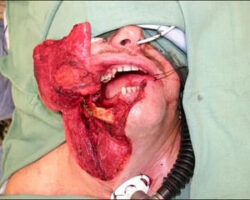
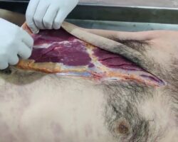
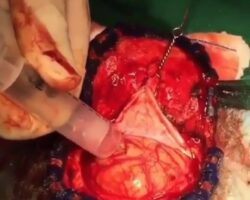
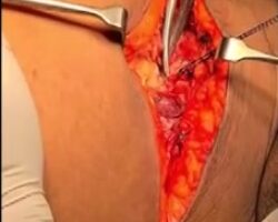
Once the surgery begins, the patient is properly positioned, and their head is secured to prevent movement. The surgeon removes a small section of the skull to access the brain. Highly precise tools and neuronavigation, which works like a GPS for the brain, are used to locate the tumor accurately. Once the brain is exposed, the surgeon removes the tumor using specialized tools such as microscopes or tweezers. If the tumor is near critical areas, the patient may remain awake to help monitor brain activity.
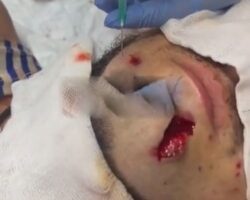
Gel foam is used to control bleeding and support healing, and it is biodegradable. After the tumor is removed, the surgeon verifies the result using imaging methods to ensure no tumor remains. The bone flap is replaced and secured. After the surgery, the patient is moved to the intensive care unit for monitoring.
Recovery typically lasts 3 to 10 days in the hospital, focusing on rest and medication to reduce swelling or prevent seizures. After discharge, the patient continues recovery at home with rest and limited activity. Rehabilitation may be needed, including physical or speech therapy if motor or cognitive functions were affected. Follow-up visits track any tumor recurrence, and additional treatments, such as chemotherapy or radiotherapy, may be recommended. Full recovery can take several months to over a year.